23 Feb X-Ray Without the Waiting Room: Precision Imaging Where and When You Need It Most
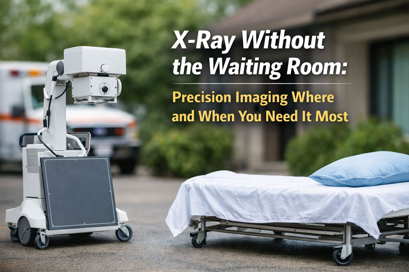
Portable x-ray: How medical imaging brings radiology to you without the waiting room
Portable x-ray: How medical imaging brings radiology to you without the waiting room is transforming the way patients access essential diagnostic x-rays. Instead of travelling to a clinic and sitting in crowded reception areas, a modern x-ray machine can now be brought directly to you. X-rays are a type of electromagnetic radiation that use radiation to create images of the inside of your body, allowing healthcare professionals to assess a broken bone, perform a chest x-ray, or examine another part of your body with precision. During an x-ray examination, x-ray beams pass through most objects, but different parts of your body absorb radiation differently depending on the density of bone and soft tissue, creating a clear x-ray image for review by a radiologist.
For patients, the x-ray procedure is straightforward and painless. You may be asked to remove jewellery or metal objects, change into a gown, or wear a lead apron to minimise radiation exposure. A technologist will position the x-ray detector or plate close to the area being x-rayed, and you might be asked to hold your breath for a few seconds while the image is taken. The amount of radiation used in modern x-ray technology is a small amount of radiation, carefully controlled to ensure safety while still delivering accurate diagnosis and treatment insights. To understand how this advanced medical imaging works in real-world settings, explore portable x-ray technology. If you need reliable, high-quality x-ray services delivered without the waiting room, now is the time to choose a mobile solution that brings radiology directly to you and supports faster, more convenient care.
What is an x-ray and how does portable x-ray technology work?
What are x-rays and how are x-rays a form of electromagnetic radiation?
Within the discussion of What is an x-ray and how does portable x-ray technology work?, it is important to understand that x-rays are a type of electromagnetic radiation. Unlike visible light, x-rays have higher energy and can pass through most objects, including the soft tissue inside of your body. This ability to use radiation to create images allows medical imaging professionals to produce pictures of the inside, helping a radiologist identify a broken bone, assess a chest x-ray, or look for signs of infection. X-rays are a form of radiation that move in beams through different parts of your body, and the resulting x-ray image shows variations depending on the density of bone, organ or soft tissue.
Although x-ray radiation sounds concerning, diagnostic x-rays involve a small amount of radiation carefully controlled during the x-ray examination. The radiation dose is considered low when compared to natural background radiation over time. During the x-ray procedure, you may be asked to remove jewellery or other metal objects, and in some cases wear a lead apron to limit radiation exposure to areas not being examined. Because x-rays are used to support diagnosis and treatment decisions, the benefit of accurate information is often far more important than the small estimated amount of radiation involved.
How does a portable x-ray machine create images compared with traditional x-ray technology?
When exploring What is an x-ray and how does portable x-ray technology work?, the next step is understanding how a portable x-ray machine operates. A modern x-ray machine generates x-ray beams that pass radiation through your body, and an image is taken as those beams are absorbed at different rates. Portable x-ray technology uses the same fundamental principles as hospital-based radiography, but the equipment is compact and designed to be transported safely. This allows a technologist to position the machine close to an x-ray area of concern, whether a bone x-ray, abdominal x-ray or x-ray of your chest, without requiring the patient to travel.
In practice, the imaging test is painless and efficient. You may be asked to stand or lie in different positions so the area being x-rayed can show up clearly. The x-ray will expose the specific part of your body needs assessment while shielding the rest. For a clearer explanation of how this equipment functions in real-world settings, see the overview of portable x-ray technology. Portable systems use radiation to create images in the same way as traditional units, ensuring reliable x-ray results while delivering radiology services directly to the patient.
What role does an x-ray detector or detector panel play in producing an x-ray image?
A key part of What is an x-ray and how does portable x-ray technology work? is the x-ray detector. The x-ray detector or plate captures the radiation that has passed through your body and converts it into a digital x-ray image. As x-ray beams travel through different parts of your body, denser structures such as bone absorb more radiation, while softer tissue allows more to pass through. The detector records these differences, enabling radiology professionals to create images of the inside with clarity and precision.
During an x-ray examination, the technologist carefully positions the x-ray detector behind the area being examined. You might be asked to hold your breath for a few seconds while the image is taken to prevent movement. The detector panel instantly transmits the pictures of the inside to a radiologist, who reviews the x-ray results for diagnosis and treatment planning. To see how this process supports patient care beyond the traditional setting, read about in-home x-rays and bedside diagnostics. This combination of modern x-ray technology and digital detection ensures high-quality medical imaging without the need for a waiting room.
Is radiation from portable x-ray safe and what is the radiation dose?
How much radiation exposure will I get from a portable x-ray compared with background radiation?
When asking, Is radiation from portable x-ray safe and what is the radiation dose?, it helps to compare x-ray exposure with everyday background radiation. X-rays are a type of electromagnetic radiation, and diagnostic x-rays use radiation to create images of the inside of your body. The amount of radiation from a single chest x-ray or bone x-ray is generally considered a small amount of radiation, often comparable to the natural background radiation you receive from the environment over a short period of time. Modern x-ray technology is designed to use the lowest radiation dose possible while still producing a clear x-ray image for accurate diagnosis and treatment.
In portable radiology, the same safety standards apply as in a hospital or clinic. The x-ray machine is carefully calibrated, and the technologist ensures that only the specific part of your body needs assessment is exposed to x-ray beams. X-rays have higher energy than visible light, allowing them to pass through most objects, but exposure is tightly controlled. For further clarification on safety considerations and common concerns, patients can review the detailed information provided in the frequently asked questions about portable x-ray services, which explains how radiation exposure is managed in real-world settings.
What is the amount of radiation in diagnostic x-rays and how is radiation dose measured?
Diagnostic x-rays are carefully measured to ensure patient safety. The radiation dose from an x-ray examination depends on the types of x-ray being performed, such as an abdominal x-ray or x-ray of your chest. Radiology professionals measure radiation dose in standardised units that reflect how much radiation is absorbed by different parts of your body. The goal is always to achieve high-quality medical imaging while keeping the amount of radiation as low as reasonably achievable. Because x-rays are used to create images that show soft tissue, bone and other structures depending on the density, a balance must be maintained between clarity and minimal exposure.
Each x-ray procedure is tailored to the individual. Factors such as age, body size and the area being x-rayed influence how the x-ray machine is set. The detector captures the radiation that passes through the body, allowing a radiologist to review the x-ray results without unnecessary repeat imaging tests. This careful approach ensures that the benefit of identifying a broken bone, signs of infection or other concerns is significantly more important than the small estimated radiation dose involved.
How can technologists reduce x-ray exposure with aprons and other precautions?
A key part of Is radiation from portable x-ray safe and what is the radiation dose? involves understanding the precautions taken during an x-ray examination. Before the image is taken, you may be asked to remove jewellery or other metal objects such as jewellery that could interfere with the x-ray image. You might also be asked to change into a gown and, in some cases, wear a lead apron. These measures help shield parts of your body that are not being examined and reduce unnecessary radiation exposure.
During the x-ray procedure, the technologist positions the x-ray detector or plate precisely behind the area being assessed. You may be asked to stand or lie in different positions and hold your breath for a few seconds while the image is taken, ensuring the x-ray shows up clearly without repeat exposure. Portable services follow the same radiography standards as fixed-site radiology, using modern equipment designed to limit radiation through your body to the smallest effective level. For more insight into how mobile radiology protects vulnerable patients while delivering diagnostic x-rays at home, see in-home imaging for vulnerable patients, which outlines how safety and patient care remain central to every examination.

What types of x-ray exams can be performed portably?
Can portable x-ray produce a chest x-ray or chest x-ray image effectively?
When considering What types of x-ray exams can be performed portably?, one of the most common questions relates to the chest x-ray. Portable radiography is highly effective for producing a clear chest x-ray image, whether to assess lung conditions, monitor infection, or investigate concerns such as fluid build-up. X-rays are a type of electromagnetic radiation that pass through soft tissue and are absorbed more readily by denser structures, allowing the x-ray image to show variations depending on the density of different parts of your body. A portable x-ray machine uses radiation to create images in exactly the same way as fixed hospital equipment, ensuring reliable medical imaging without compromising diagnostic quality.
In practice, the x-ray examination is straightforward and painless. The technologist positions the x-ray detector or plate behind the patient’s chest, and you may be asked to hold your breath for a few seconds while the image is taken. This helps the x-ray shows the lungs and surrounding structures clearly. For patients unable to travel, this approach allows radiology services to deliver essential chest imaging safely and efficiently in a home or care setting. More detail about how these services operate can be found on the mobile x-ray services overview, which explains how portable systems maintain high clinical standards.
Are bone x-ray and broken bone assessments possible with portable radiography?
Another key area within What types of x-ray exams can be performed portably? involves bone x-ray assessments. Portable x-ray technology is well suited to diagnosing a broken bone or evaluating joint injuries. Because x-rays have higher energy than visible light, x-ray beams pass through most objects but are absorbed strongly by bone. This contrast enables the x-ray image to show fractures, alignment issues and other abnormalities clearly. Diagnostic x-rays of the wrist, hip, ankle or other parts of your body can be completed on-site, reducing the need for transport after an injury.
During the x-ray procedure, the area being x-rayed is positioned carefully, and you may be asked to stand or lie in different positions so the image can show up clearly. The amount of radiation used remains a small amount of radiation, controlled to ensure safety while achieving accurate x-ray results. The detector captures detailed pictures of the inside of the affected region, which a radiologist reviews to support diagnosis and treatment planning. This makes portable radiography a practical and clinically sound option for musculoskeletal assessments outside a traditional clinic.
Can portable imaging perform abdominal x-ray or other imaging tests like dental x-ray?
Within the scope of What types of x-ray exams can be performed portably?, abdominal x-ray examinations are also possible in many cases. An abdominal x-ray can help assess the digestive system, detect kidney stones, or investigate pain in a specific part of your body. The portable x-ray machine uses radiation to create images of the inside, and the x-ray detector records how electromagnetic radiation passes through different tissues. As with other types of x-ray, the image is taken quickly, and the process remains painless.
Portable services focus solely on x-ray and do not provide ultrasound, CT or MRI. While a dental x-ray typically requires specialised dental equipment, many general diagnostic x-rays — including chest, limb and certain abdominal studies — can be performed safely with portable radiography. Patients may be asked to remove jewellery or metal objects and, in some situations, wear a lead apron to minimise radiation exposure. For further insight into how in-home radiology supports those who cannot attend a facility, see how portable imaging is changing lives, which outlines how these imaging tests enhance access to timely medical imaging.
How is an x-ray examination performed at bedside and what may be asked?
What happens during a portable x-ray procedure and will it be painless?
Understanding How is an x-ray examination performed at bedside and what may be asked? begins with knowing what to expect during the x-ray procedure itself. A portable x-ray machine is brought directly to the bedside, where a technologist positions the equipment safely around the patient. X-rays are a type of electromagnetic radiation used to create images of the inside of your body, and the process is generally painless. The x-ray beams pass through different parts of your body, and depending on the density of bone and soft tissue, the x-ray image will show areas that absorb more or less radiation. Whether it is a chest x-ray, bone x-ray or another imaging test, the goal is to obtain clear pictures of the inside without causing discomfort.
In most cases, the examination takes only a few minutes. The technologist places the x-ray detector or plate behind the area being x-rayed, and the image is taken while you remain still. You may be asked to hold your breath for a few seconds so the x-ray shows up clearly. The amount of radiation used in modern x-ray technology is carefully controlled, and the radiation dose remains low while still allowing accurate diagnosis and treatment decisions. For further details about how this process works in practical settings, see the explanation of x-ray on demand at the bedside.
What may be asked about jewellery, clothing, or positioning before the x-ray examination?
Before the x-ray examination begins, you may be asked to remove jewellery or other metal objects such as jewellery that could interfere with the x-ray image. Metal can block x-ray beams and affect how the detector records the radiation passing through your body. Depending on the part of your body being examined, you might be asked to change into a gown to ensure clothing does not obscure the area being x-rayed. These steps are standard in radiography and help produce accurate diagnostic x-rays.
Positioning is also important. You may be asked to stand or lie in different positions so the x-ray will expose the correct angle of the part of your body needs assessment. For a chest x-ray, you might be asked to hold your breath briefly; for other types of x-ray, you may need to remain still while the image is taken. In some cases, you may be asked to wear a lead apron to protect areas not being examined, further reducing radiation exposure. These precautions ensure that the small amount of radiation used is focused only where necessary.
Who performs the bedside x-ray — a radiologist or technologist — and how do they create images?
A bedside x-ray is typically performed by a trained technologist who specialises in radiography. The technologist operates the x-ray machine and positions the x-ray detector carefully to create images of the inside of your body. After the image is taken, the x-ray results are reviewed by a radiologist, a medical specialist in radiology who interprets the x-ray image and provides a report to the healthcare provider. This collaborative approach ensures both technical accuracy and expert diagnostic interpretation.
During the process, the technologist ensures correct placement of the detector and controls the radiation exposure so that only the intended area is examined. The x-rays are used to create images that can reveal a broken bone, changes in soft tissue, or other abnormalities. Once captured, the digital images are transmitted securely for review. To understand more about how bedside medical imaging supports patients who cannot travel, read about in-home x-rays and bedside diagnostics, which outlines how radiology services are delivered safely and effectively outside a traditional clinic environment.

When will I get x-ray results and what do x-ray results show?
How long until x-ray results are available and will a radiologist interpret the x-ray image?
When considering When will I get x-ray results and what do x-ray results show?, timing is often a key concern. In most cases, once the x-ray examination is complete and the image is taken, the digital x-ray image is transmitted securely for review. A radiologist, who specialises in radiology and medical imaging, interprets the diagnostic x-rays and prepares a report for the healthcare provider. Because modern x-ray technology uses advanced digital detectors, images are available almost immediately after the x-ray procedure, significantly reducing delays compared with older film-based radiography.
Although the image is captured within minutes, the final x-ray results depend on reporting workflows and clinical urgency. Urgent chest x-ray or bone x-ray findings may be prioritised to support rapid diagnosis and treatment. The radiologist reviews how x-ray beams have passed through different parts of your body, assessing variations depending on the density of bone and soft tissue. For a broader understanding of how portable reporting integrates with clinical care, see the overview of portable radiology and diagnostic imaging services, which explains how images move from bedside to interpretation.
What can x-rays show—soft tissue, broken bone, kidney stones, or inside of your body?
A common question within When will I get x-ray results and what do x-ray results show? is what exactly an x-ray can detect. X-rays are a type of electromagnetic radiation used to create images of the inside of your body. Because x-rays have higher energy than visible light, they pass through most objects but are absorbed differently by bone and soft tissue. This allows an x-ray image to clearly identify a broken bone, changes in joint alignment, or certain abnormalities in the chest. An abdominal x-ray may help detect kidney stones or signs of obstruction in the digestive system.
While x-rays can also show some soft tissue changes, they are most effective for assessing bone and air-filled structures such as the lungs. The x-ray shows contrast based on how radiation passes through the area being x-rayed, producing pictures of the inside that assist with diagnosis and treatment planning. During the x-ray examination, the technologist ensures the correct positioning so the part of your body needs assessment is accurately captured. This targeted approach keeps radiation exposure low while still delivering valuable diagnostic information.
How do modern x-ray images and x-ray technology compare in clarity and diagnostic value?
Modern x-ray technology has significantly improved the clarity and diagnostic value of x-ray images. Digital x-ray machines use advanced detectors to capture radiation through your body and instantly convert it into high-resolution images. Unlike older systems, today’s equipment allows radiology professionals to adjust contrast and brightness without repeating the x-ray procedure. This reduces unnecessary x-ray exposure and enhances the ability to identify subtle findings in bone, lung tissue or other structures.
High-quality diagnostic x-rays rely on precise positioning, correct radiation dose, and advanced detector sensitivity. The amount of radiation used remains a small amount of radiation, carefully calibrated to achieve optimal image quality. The combination of digital radiography and experienced interpretation ensures that x-ray results provide reliable insight into the condition being investigated. For further explanation of how bedside imaging delivers consistent clarity outside a hospital environment, explore in-home portable x-ray services, which outlines how modern systems maintain diagnostic standards while offering greater convenience.
Conclusion
In conclusion, understanding how x-ray technology works — and how portable radiology delivers it safely — helps patients feel confident about the process. X-rays are a type of electromagnetic radiation used to create images of the inside of your body, allowing a radiologist to assess a broken bone, review a chest x-ray, or investigate concerns identified during a medical imaging assessment. A modern x-ray machine uses controlled radiation to create images that show differences in soft tissue and bone depending on the density of each structure. The amount of radiation involved in diagnostic x-rays is a small amount of radiation, carefully managed to minimise radiation exposure while still producing clear and reliable x-ray results. From positioning the x-ray detector or plate to ensuring the correct part of your body needs evaluation, each x-ray examination follows strict radiography standards.
Portable X-Ray Services extend this capability beyond the traditional clinic, bringing high-quality x-ray examinations directly to the bedside. The x-ray procedure remains painless, and patients may be asked to remove jewellery, change into a gown, or wear a lead apron as a precaution. The image is taken within seconds, and the results are reviewed by a radiologist to support accurate diagnosis and treatment decisions. To learn more about how this works in practice, explore booking a portable x-ray service at your door or read about bringing diagnostics to your doorstep. If you require dependable, professional x-ray services delivered without the waiting room, choose a provider that combines advanced x-ray technology with responsive patient care and clinical expertise.